Product Highlights
VERIQA
Patient QA Platform
RUBY
Modular QA Phantoms
BEAMSCAN
Water Phantom
BEAMSCAN MR
Water Phantom for MRgRT
OCTAVIUS 4D / 4D MR
QA Phantom
Electron Density Phantom
Classical Shape Phantom
Track-it
QA Data Management Platform
flashDiamond
Detector
UNIDOS Tango & Romeo
Reference Class Electrometers
All Radiation Therapy Products
A B C
D
E F
G
H
I
J
K
L
M
N
O P Q R S T U V W
X
Y
Z
Product Highlights
NOMEX
Multimeter
NOMEX
Dosemeter
Track-it
QA Data Management Platform
QRM
Phantoms
All Diagnostic Imaging Products
A B C
D
E
F
G
H
I
J
K
L M N
O
P
Q
R S T U
V
W
X
Y
Z

FLASH Radiotherapy:
Dosimetric Challenges and Solutions

Sparing of healthy tissue while maintaining or improving tumor control is the primary goal of radiotherapy. In recent decades, this goal has been advanced primarily through technological innovations. Since the rediscovery of the so-called FLASH effect in 2014, many preclinical studies have shown that ultra-high dose rate (UHDR) irradiation (mean dose rate > 40 Gy/s) results in significant healthy tissue sparing. Accurate dosimetry of these ultra-high dose rates is a major challenge and crucial for the safe transition of FLASH radiotherapy (FLASH-RT) into clinical practice. To address this need, PTW had joined the European UHDpulse project to provide a framework for dose measurements at UHDR1, which was successfully completed in February 2023.


Ionization chambers need to be adapted to operate under high dose rate conditions

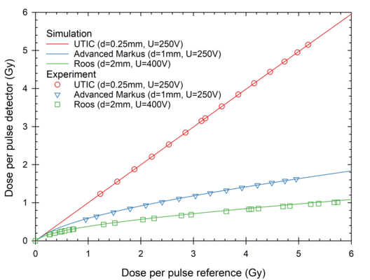
Vented ionization chambers (ICs) are considered as the gold standard for reference dosimetry and are most commonly used as secondary standards. Unfortunately, established ICs show significant saturation effects due to ion recombination at UHDR in pulsed beams (Figure 1).


Figure 1: Detector reading from parallel plate ionization chambers with different electrode distances d and operational voltages U (Roos PTW T34001 (d=2mm), Advanced Markus PTW T34045 (d=1mm) and a prototype PTW UTIC (d=0.25 mm)) without correction for ion recombination effects as function of dose per pulse from experiment (symbols) and simulations (lines)

In addition, other conventional active real-time detectors, such as solid-state detectors based on semiconductors, start to fail at UHDR. For this reason, passive dosimeters, such as alanine, radiochromic films, or luminescence dosimeters (TLDs and OSLDs), are mainly used under these conditions. However, the use of passive dosimeters is complex, very time-consuming, and usually lacks the traceability to primary standards. There is therefore a need for active real-time dosimeters and traceability. It is also desirable to use ionization chambers as a secondary standard under UHDR conditions.

To find a solution to this challenge, the role of the distance between the electrodes of vented ICs and its impact on ion recombination has been analyzed experimentally and by numerical simulation2. The results show that this parameter is the most relevant to achieve negligible recombination losses. Using a very small electrode distance of 0.25 mm in vented ICs (called ultra-thin ICs (UTIC)) leads to a charge collection efficiency higher than 99% for a 2.5 µs pulse of 5 Gy (Figure 1). Ionization chambers are therefore excellent candidates as secondary standard for reference dosimetry in UHDR beams because they are also waterproof, easy to handle and can be used according to the existing methodology in current codes of practice.


flashDiamond – the first  detector for FLASH dosimetry

However, ICs are limited in spatial resolution due to the diameter of the sensitive volume, especially when measuring lateral dose distributions and in small fields.

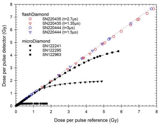
A detector that is particularly well suited for these applications, which require high spatial resolution, is the well-established microDiamond detector by PTW (type T60019).It can also be used for electron radiation, including high dose per pulse (DPP) applications such as used in IOeRT3,4.  Due to the high water equivalence of its sensitive volume in terms of effective atomic number, no conversion from ion dose to absorbed dose to water is necessary when determining depth dose curves for electron beams. This characteristic, along with its good stability of response as a function of the accumulated dose, made the microDiamond detector a promising detector for use in UHDR1 environments. However, the detector has shown to exhibit saturation behavior at UHDR pulsed electron beams5. A thorough investigation under ultra-high pulse dose rate (UHPDR) conditions using different samples and modified designs was performed by Marinelli et al.6 and Kranzer et al.7. These studies confirmed the observed saturation behavior (Figure 2) and investigated the influencing parameters.

Figure 2: Detector reading as function of the dose per pulse in a water phantom for three microDiamond detectors (PTW T60019) under the same beam conditions (2.5 µs pulse duration) as well as for two flashDiamond detectors (PTW T60025) at different pulse durations t

It was found that the critical parameters to improve the dose-response linearity of the detector are the sensitivity and the total series resistance. Optimizing these parameters results in a new type of detector adapted to UHPDR conditions, the flashDiamond detector by PTW (type T60025). This new detector, which is now commercially available, has been successfully tested in terms of linearity with UHDPDR (Figure 2), three-dimensional relative dose distributions (percentage depth dose curves and lateral dose profiles) and output factors8.

Together with the flashAdapter (T16055), the flashDiamond detector can be used with PTW electrometers and integrated into scanning water phantoms, which makes the application very easy and familiar for medical physicists. The flashDiamond is therefore an ideal detector for a wide range of applications under UHDR conditions.

Overview on PTW FLASH dosimetry solutions available for preclinical use

    References

    • Schüller, A. et al. The European Joint Research Project UHDpulse – Metrology for advanced radiotherapy using particle beams with ultra-high pulse dose rates. Phys. Medica 80, 134–150 (2020).
    • Kranzer, R. et al. Charge collection efficiency, underlying recombination mechanisms, and the role of electrode distance of vented ionization chambers under ultra-high dose-per-pulse conditions. Phys. Medica 104, 10–17 (2022).
    • Di Venanzio, C. et al. Characterization of a microDiamond detector in high-dose-per-pulse electron beams for intra operative radiation therapy. Phys. Medica 31, 897–902 (2015).
    • Güngör, G., Aydın, G., Mustafayev, T. Z. & Özyar, E. Output factors of ionization chambers and solid state detectors for mobile intraoperative radiotherapy (IORT) accelerator electron beams. J. Appl. Clin. Med. Phys. 20, 13–23 (2019).
    • Di Martino, F. et al. FLASH Radiotherapy With Electrons: Issues Related to the Production, Monitoring, and Dosimetric Characterization of the Beam. Front. Phys. 8:570697  (2020). doi: 10.3389/fphy.2020.570697.
    • Marinelli, M. et al. Design, realization and characterization of a novel diamond detector prototype for flash radiotherapy dosimetry. Med. Phys. (2022). doi:10.1002/mp.15473
    • Kranzer, R. et al. Response of diamond detectors in ultra-high dose-per-pulse electron beams for dosimetry at FLASH radiotherapy. Phys. Med. Biol. 67, 075002 (2022).
    • Rinati, G. V. et al. Application of a novel diamond detector for commissioning of FLASH radiotherapy electron beams. Med. Phys. (2022). doi:10.1002/mp.15782

    About the author

    Rafael Kranzer studied biomedical engineering at Technische Hochschule Mittelhessen (THM) and completed his doctorate at Carl von Ossietzky University. He works as Team Lead Radiation Physics at PTW Freiburg, focusing on research and development of dosimetry equipment and detectors.

    Diese Seite existiert auch in Ihrer Sprache.
    This page also exists in your language.
    Sprache wechseln Change Language